Epithelium off Cornea collagen cross linking (CXL) is not voodoo. We realize that early in new technology a lot of unfounded claims like epi on CXL being Better than epi off might be made. It behooves us clinicians to present proof to peers.
There is a symbiosis between diagnostic technology and treatments. We will take help of Carl Zeiss Ocular coherence tomography to demonstrate the effectiveness of epi off CXL.
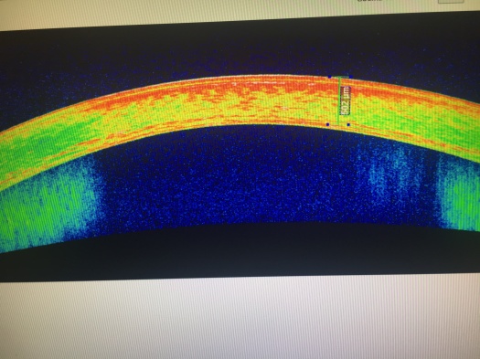
Here is a High Density picture of a keratoconus cornea before any treatment. It is early keratoconus in a young girl. This is the best time to intervene to treat Keratoconus. Look how uniform the picture is. The top layer of epithelium can be clearly seen. We remove this layer with laser or as Professor Theo Seiler recommends with ethyl alcohol.
Epithelium prevents riboflavin from entering the stroma and hinders the UV absorption. Look at the picture below. You can clearly see a haze which ends around 80 % depth as a line of demarcation.
The same line of demarcation after cornea cross linking is better highlighted in they colored OCT of the Cornea.
If you are suffering from Keratoconus Call 805-283-6520 to see if cross linking of cornea is the best option for you.
Update terbaru menghadirkan map baru yang luas, lanjut ke ulasan Keluaran macau. Update terbaru memperbaiki sistem matchmaking. Pertandingan jadi terasa lebih seimbang.
Slot Gacor Online menawarkan berbagai variasi permainan yang cocok untuk semua jenis pemain. Mulai dari yang suka permainan cepat hingga yang menikmati strategi, semuanya tersedia di sini.
Kombinasi simbol dalam permainan ini sangat menentukan keberhasilan pemain dalam meraih kemenangan besar. Pemahaman terhadap mekanisme permainan akan sangat membantu dalam menciptakan strategi yang lebih efektif. Tidak heran jika semakin banyak orang yang tertarik untuk memainkan Mahjong Ways 2.
Promo top-up ini sangat cocok untuk kamu yang ingin mendapatkan resource lebih banyak dengan harga lebih hemat lihat informasinya pedetogel link alternatif. Tips leveling cepat bisa dilakukan dengan konsisten menyelesaikan misi harian. Cara ini terbukti cukup efektif.
Cara cepat naik tier tanpa harus main nonstop ternyata ada triknya sendiri, tutorial lengkap di bd-innovations.com. Banyak pemain mencari setting grafis terbaik untuk FPS stabil. Performa lancar memang penting saat pertandingan kompetitif.
Event mini game dalam game utama ini ternyata kasih reward utama kalau target tercapai, cara mainnya dijelaskan pada toto. Tips farming efektif adalah pilih map dengan respawn cepat. Efisiensi waktu sangat berpengaruh.
Deposit Kecil Membawa Pengalaman Bermain Slot Tanpa Mengeluarkan Dana Besar
Bermain slot online menjadi lebih hemat dengan adanya Slot Depo 5k. Bagi pemain baru yang masih ingin mencoba-coba, deposit kecil ini memungkinkan mereka untuk merasakan pengalaman bermain tanpa harus mengeluarkan dana besar. Selain itu, fleksibilitas dalam deposit memberikan pemain lebih banyak opsi. Slot Depo 5k benar-benar membawa suasana bermain slot yang ramah di kantong.
Promo top-up hari ini cocok buat stok resource jangka panjang tanpa harus nunggu event besar di toto. Update visual lingkungan bikin map terasa lebih hidup. Detail kecil bikin betah.
Event terbatas ini jadi momen terbaik buat farming item langka dalam Toto. Event komunitas sering menghadirkan hadiah kecil tapi menarik. Partisipasi pemain membuat suasana semakin meriah.
Bukan rahasia lagi, kalau rtp live jadi alat wajib para pemain slot buat nyari slot rtp tertinggi yang paling gacor. Dengan info ini, kamu nggak cuma asal main, tapi bisa pilih slot gacor hari ini yang beneran ngasih peluang besar. Sensasinya beda banget dibanding main asal nebak, tiap spin jadi lebih berarti dan bikin kamu nggak sabar buat putaran berikutnya.
Jika kamu ingin mencoba peruntungan tanpa beban besar, pilihan terbaik adalah Slot 10k yang memberikan kesempatan bermain lebih lama dengan dana yang minim.
Bermain dengan nyaman dan lancar kini menjadi lebih mudah ketika memilih slot server Thailand sebagai pilihan utama, karena kecepatan dan kestabilannya memastikan pengalaman bermain yang menyenangkan tanpa gangguan yang mengurangi fokus dalam meraih kemenangan besar.
Live Draw Macau Transparan Memberikan Kepercayaan Penuh
Melihat angka keluar secara langsung melalui Live draw macau menghadirkan kepercayaan penuh, karena pemain bisa menyaksikan sendiri proses pengundian yang berlangsung terbuka.
Saat fokus mencari kenyamanan dan keamanan, para pemain mengandalkan Situs Togel yang menyediakan berbagai pasaran lengkap, transaksi cepat dan aman, serta informasi angka keluar terbaru yang membantu mereka merencanakan strategi bermain lebih efektif.
Banyak penggemar judi online memastikan taruhan mereka ditempatkan di Togel Resmi yang diawasi secara ketat agar semua hasil angka keluaran valid, transaksi aman, dan hadiah dibayarkan tepat waktu.
Promo top-up dengan bonus item langka ini cuma tersedia hari ini, ketentuannya dijelaskan pada togel. Update fitur sosial bikin interaksi antar pemain makin gampang. Mulai dari chat sampai party system.
Promo top-up terbatas ini ideal buat persiapan turnamen di situs toto. Event spesial weekend biasanya lebih santai. Cocok buat main bareng teman.
Leveling cepat buat karakter kedua bisa lebih gampang dengan metode situs toto. Komunitas biasanya punya istilah sendiri. Pemain baru perlu waktu buat ngerti.
Promo top-up plus voucher lagi dibuka, klaim bonusnya lewat Togel. Update ranking system kadang bikin progress terasa lebih berat. Tetap konsisten, lama-lama naik juga.
Patch baru bawa item yang bikin farming makin cepat, tapi banyak yang belum tau cara pakainya, cek di toto. Event seasonal bikin suasana game berubah total. Map dan musiknya biasanya beda dari biasanya.
Promo top-up ini kasih bonus diamond tambahan kalau beli di jam tertentu, jamnya ada di togel. Event battle pass itu paling efektif kalau kamu konsisten. Sedikit tiap hari lebih ringan daripada kebut di akhir.
Penjelasan Guru Sosiologi Tentang Peluang Hidup
Seorang guru sosiologi menjelaskan contoh perilaku sosial kepada muridnya, lalu di tengah penjelasan ia menyinggung Togel hanya sebagai ilustrasi tentang cara manusia memahami peluang dan harapan.
Turnamen guild yang paling ditunggu pemain akhirnya segera dimulai, jadwal pertandingannya bisa kamu cek pada Togel Online. Event spesial kadang memberikan bonus exp yang besar. Hal ini membantu pemain naik level lebih cepat.
Dengan layanan pelanggan yang siap 24 jam, Situs Togel178 memastikan bahwa setiap pertanyaan dan masalah pemain dapat diatasi dengan cepat dan efektif, memberikan pengalaman bermain yang lebih nyaman.
Dengan layanan pelanggan yang siap 24 jam, Togel178 memastikan bahwa setiap pertanyaan dan masalah pemain dapat diatasi dengan cepat dan efektif, memberikan pengalaman bermain yang lebih nyaman dan bebas dari masalah.
Promo bonus top-up terbatas malam ini bikin player ramai masuk, lengkapnya tautan arah pedetogel alternatif. Patch baru kadang buff karakter lama yang sempat dilupakan pemain. Itu bikin variasi pilihan makin luas.
Tips push exp dan resource sekaligus mulai dicoba, tutorial arah rujukan hk pools 20. Event double drop selalu jadi waktu favorit buat grinding. Hasil farming biasanya jauh lebih maksimal saat itu.
Partner Links
Petaruh www.resea-rchgate.net Singapore mengandalkan data lengkap SGP untuk pasang angka jitu mereka.
Para pemain toto di sini Rtp Togel178 takkan suntuk, beragam pasaran togel siap dimainkan.
Bonus deposit Togel 279 sangat menguntungkan bagi pemain poker online.
Cara Togel158 bermain yang fleksibel membantu pemain bergabung ke situs judi terpercaya.
Kami siapkan analisis tenis lengkap dengan statistik Togel158 pertandingan, rekor head-to-head, dan performa terkini.
Cara termudah untuk menerapkan Togel178 spekulasi hebat ini agar pemain merasakan servis yang lebih baik.
Situs ini tidak cuma tempat bermain, tapi juga sumber panduan bermanfaat dan macauindo.co mendalam.
Strategi tidak selalu https://pedetogel.net/ sempurna, tetapi dengan latihan terus, keterampilanmu akan tajam.
Di sini, kami akan bagikan panduan sukses menang Togel178 dalam permainan Kompetisi online terbaik.
Ini telah jadi faktor utama Situs Pedetogel Rekreasi online sukses sampai sekarang.
Registrasi simpel dan cepat, dimulai main sabung https://kampuspoker.com/ ayam online dengan cepat.
Akhirnya, kami situs togel ingin menegaskan pentingnya sabar dan disiplin dalam bermain Aktivitas Seru secara daring.
Dengan cara ini, Slot Gacor Anda bisa tingkatkan keuntungan dan sukses dalam perjudian.
Berkumpul dengan teman di tempat nongkrong bisa kaya situs idn poker lewat permainan 1 tanpa bekerja.
Banyak pemain togel memilih Colok178 sebagai situs andalan mereka. Selain memiliki reputasi terpercaya, Colok178 menawarkan kemudahan transaksi melalui berbagai metode, termasuk e-wallet dan bank lokal. Dengan peluang menang yang tinggi, situs ini menjadi favorit banyak kalangan pencinta togel.
Banyak bettor mencari situs yang tidak hanya aman tetapi juga memberikan banyak keuntungan tambahan. Pedetogel menawarkan berbagai promo menarik yang dapat meningkatkan peluang kemenangan pemain. Dari bonus deposit hingga cashback, semuanya tersedia bagi para pengguna setia. Selain itu, situs ini memiliki sistem transaksi yang sangat cepat dan fleksibel, memungkinkan pemain untuk menikmati permainan tanpa hambatan. Dengan berbagai keunggulan ini, tidak heran jika situs ini semakin diminati.
Bagi pemain yang ingin mendapatkan hasil terbaik, situs macau menawarkan berbagai pilihan bonus dan promosi yang terus diperbarui. Pemain bisa memanfaatkan kesempatan ini untuk meningkatkan peluang menang mereka di setiap taruhan.
Bermain togel online bukan hanya soal keberuntungan tetapi juga soal memilih platform yang terpercaya. Salah satu cara memastikan keamanan bermain adalah dengan bergabung di situs togel resmi yang memiliki reputasi baik. Situs ini menggunakan sistem keamanan tinggi dan pembayaran yang cepat untuk menjamin kenyamanan pemain. Selain itu, berbagai fitur tambahan seperti statistik angka dan analisis tren juga disediakan untuk membantu pemain meningkatkan peluang menang.
Salah satu fitur menarik yang kini populer di kalangan pemain adalah cashback mingguan. Bonus ini memungkinkan pengguna untuk mendapatkan kembali sebagian dari kekalahan mereka. Tidak heran jika Toto92 menjadi favorit karena rutin memberikan cashback dan promosi lainnya.
Dalam memilih situs permainan online, penting untuk memperhatikan reputasi serta pengalaman yang dimiliki oleh platform tersebut. Toto92 sudah lama dikenal sebagai salah satu pionir dalam industri ini. Dengan pengalaman bertahun-tahun, mereka paham apa yang dibutuhkan pemain dan selalu berupaya memberikan yang terbaik. Hal ini menjadi alasan utama banyak pemain tetap setia.
Salah satu hal yang membuat platform hiburan digital berkembang pesat adalah kepercayaan pengguna. Nama Sabatoto muncul sebagai pilihan yang konsisten menjaga kualitas dan memberikan pelayanan terbaik. Hal ini dibuktikan dari banyaknya ulasan positif yang beredar.
Platform yang ramah pengguna biasanya mendapat tempat di hati masyarakat. Hal inilah yang menjadi fokus dari Pedetogel, menciptakan pengalaman tanpa ribet untuk semua kalangan.
Menjadi bagian dari komunitas Togel178 memberikan keuntungan tersendiri karena ada banyak tips dan strategi dari para pemain senior.
Karena sering kesulitan klaim bonus di situs lain, saya akhirnya pindah ke Pedetogel dan ternyata di sini semua promosi bisa diklaim otomatis tanpa perlu menghubungi admin atau repot isi form.
Layanan aplikasi mobile yang disediakan oleh Togel158 membuat pengalaman bertaruh semakin fleksibel dan praktis, karena pemain bisa mengakses permainan kapan pun dan di mana pun tanpa batasan perangkat.
Tidak hanya mengutamakan kecepatan transaksi, Sabatoto juga menjamin semua pasaran yang tersedia adalah resmi dengan hasil angka yang selalu transparan dan akurat.
Sistem notifikasi otomatis di Togel178 memberi info update cepat sehingga di tengah jadwal padat, pemain tidak ketinggalan hasil draw atau promo terbaru dari situs.
Dalam urusan pembayaran, Togel178 selalu memproses penarikan dengan cepat sehingga pemain tidak perlu menunggu lama untuk menikmati hasil kemenangan mereka.
Deposit melalui e-wallet, pulsa, maupun transfer bank bisa dilakukan dengan mudah di Pedetogel sehingga pemain memiliki banyak pilihan transaksi sesuai kebutuhan.
Pemain yang aktif setiap hari berkesempatan mendapatkan reward tambahan dari Pedetogel melalui program loyalitas khusus.
Bagi para pemain lama, Sabatoto menjadi tempat favorit karena selain lengkap, mereka juga konsisten membayar kemenangan tepat waktu.
Banyak promo cashback dihadirkan oleh Sabatoto sebagai bentuk apresiasi untuk para member setia yang terus bermain dan menikmati setiap pasaran yang tersedia.
Bagi yang baru ingin mencoba judi togel online, Togel279 menyediakan panduan lengkap yang membantu pemula memahami cara taruhan hingga teknik prediksi angka dengan mudah.
Para penggemar togel sangat puas dengan layanan dari Togel279 yang selalu memberikan update angka keluaran secara real-time, sehingga tidak perlu menunggu lama untuk mengecek hasil.
Semua pemain yang bergabung di Togel158 dapat menikmati fasilitas discount dan bonus yang membuat setiap taruhan jadi lebih hemat dan berpeluang meraih kemenangan besar.
Togel158 memastikan setiap proses pendaftaran member baru berjalan cepat dan tanpa kendala, sehingga siapapun dapat langsung menikmati berbagai layanan unggulan yang tersedia di platform ini.
Tidak hanya itu, Colok178 juga menawarkan berbagai fitur tambahan yang dirancang khusus untuk meningkatkan kenyamanan pengguna dan mendukung permainan yang lebih seru.
Banyak orang memilih untuk bermain di Togel279 karena reputasinya yang terpercaya serta dukungan layanan yang responsif setiap saat tanpa henti.
Banyak bettor yang menilai keberhasilan mereka dalam taruhan meningkat setelah menggunakan berbagai tools analitik dan statistik pertandingan yang disediakan secara lengkap di Sbobet88.
Bagi mereka yang sudah berpengalaman, bermain dengan slot bet 200 memungkinkan eksplorasi bonus besar dan fitur premium, memberikan rasa puas yang berbeda dibanding taruhan minimal.
Proses pendaftaran di agen permainan resmi sangat sederhana karena alurnya sudah dioptimalkan melalui dukungan Pedetogel sehingga pemain dapat bergabung cepat.
Proses pendaftaran pada agen resmi berjalan lancar dibantu integrasi Togel279 yang membuat seluruh tahapan lebih mudah dipahami.
Proses pendaftaran pada agen hiburan angka resmi yang terhubung Togel Resmi dibuat sederhana agar mudah diakses semua kalangan.
Proses pendaftaran pada layanan resmi dibuat praktis karena Bandar Togel mengutamakan efisiensi tanpa mengurangi keamanan akun.