Epithelium off Cornea collagen cross linking (CXL) is not voodoo. We realize that early in new technology a lot of unfounded claims like epi on CXL being Better than epi off might be made. It behooves us clinicians to present proof to peers.
There is a symbiosis between diagnostic technology and treatments. We will take help of Carl Zeiss Ocular coherence tomography to demonstrate the effectiveness of epi off CXL.
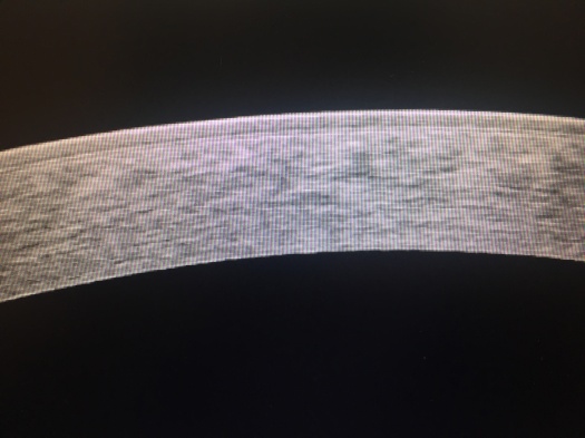
Here is a High Density picture of a keratoconus cornea before any treatment. It is early keratoconus in a young girl. This is the best time to intervene to treat Keratoconus. Look how uniform the picture is. The top layer of epithelium can be clearly seen. We remove this layer with laser or as Professor Theo Seiler recommends with ethyl alcohol.

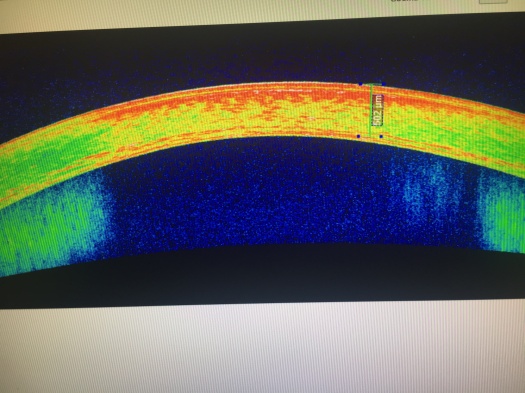
Epithelium prevents riboflavin from entering the stroma and hinders the UV absorption. Look at the picture below. You can clearly see a haze which ends around 80 % depth as a line of demarcation.

The same line of demarcation after cornea cross linking is better highlighted in they colored OCT of the Cornea.

If you are suffering from Keratoconus Call 805-283-6520 to see if cross linking of cornea is the best option for you.

